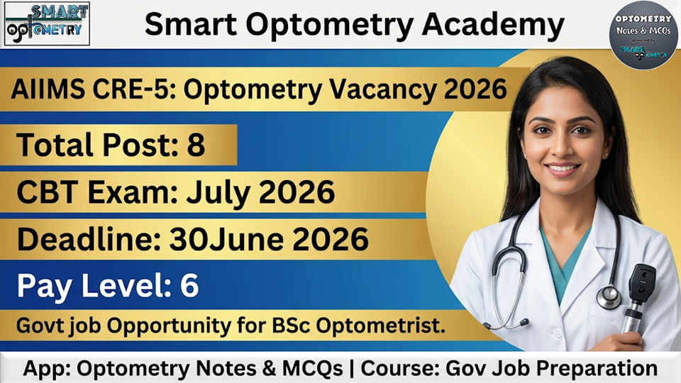
📋 DSSSB Optometrist 2026 · Post Code 09/26
📄 2019 Paper
DSSSB Optometrist
Previous Year Paper 2019
Part 4 — Q61 to Q80
20 actual questions from DSSSB Optometrist 2019 covering Retinoscopy, Diabetic Retinopathy, Retinal Detachment, 3rd Nerve Palsy, OCT, LogMAR, Strabismus, Eye Bank, Vision 2020, Slit-Lamp & Community Ophthalmology — click any option to reveal the answer instantly.
📅 Exam Year: 2019
❓ Questions: Q61–Q80
📌 Vacancies 2026: 15 Posts
⏰ Last Date: 28 Mar 2026
This blog contains questions Q61 to Q80 from the DSSSB Optometrist Previous Year Question 2019 with correct answers and detailed explanations. Topics covered: Retinoscopy essentials, Diabetic Macular Edema, Retinal Detachment layers, 3rd Nerve Palsy vs Myasthenia Gravis, Neonatal Conjunctivitis, Suture types, Cataract surgery contraindications, LogMAR scale, OCT imaging, Double Maddox Rod, Prism Cover Test, Hand Hygiene, Low Vision classification, Global Blindness causes, Eye Care Camp Teams, Vision 2020 launch, Eye Bank functions, and Slit-Lamp limitations.
How to use: Click your chosen option → the correct answer and full explanation appear immediately. Track your score with the floating card (drag anywhere on screen).
📑 Questions in This Part (Q61–Q76)
Answer your first question to begin →
ARetinoscope
BOphthalmoscope
CRed and green filter
DSnellen’s chart
👆 Click an option to check your answer
✓ Correct Answer: A. Retinoscope
Retinoscopy is an objective technique for determining refractive error by observing the movement of the red reflex from the patient’s retina. The retinoscope is the absolute, indispensable instrument — it projects a streak of light into the eye and allows the examiner to observe the direction and speed of the reflex movement. No other instrument can substitute it for performing retinoscopy. Snellen’s chart is used for subjective acuity testing, not retinoscopy.
AMicroaneurysm
BMacular edema
CRetinal hemorrhage
DRetinal detachment
👆 Click an option to check your answer
✓ Correct Answer: B. Macular edema
Diabetic Macular Edema (DME) is the leading cause of moderate-to-severe vision loss in diabetic retinopathy. Breakdown of the blood-retinal barrier causes fluid and lipid exudates to leak directly into the macula, disrupting central photoreceptor function. Microaneurysms and retinal hemorrhages are classic early signs but often in the peripheral retina — they do not directly affect central vision unless they occur near the fovea. Retinal detachment is a late, serious complication but less common.
ARPE and Bruch’s membrane
BNerve fibre layer and rest of retina
COuter plexiform layer and inner nuclear layer
DNeurosensory retina and retinal pigment epithelium
👆 Click an option to check your answer
✓ Correct Answer: D
Retinal detachment occurs when subretinal fluid (SRF) accumulates in the potential space between the neurosensory retina and the RPE. This separates the photoreceptor outer segments from the RPE which normally provides metabolic support, oxygen, and removes photoproducts. Without this contact → rapid photoreceptor degeneration and irreversible vision loss if not treated promptly. Note: the embryological origin of this space is the optic cup lumen — the neurosensory retina and RPE are only loosely attached by interdigitation, not tight junctions.
APtosis
BIpsilateral mydriasis
CPtosis increases in the evening
DDefective accommodation
👆 Click an option to check your answer
✓ Correct Answer: C
Ptosis that worsens in the evening is a classic sign of Myasthenia Gravis (fatigable ptosis — worsens with sustained use), NOT 3rd nerve palsy. Signs of 3rd CN palsy: Complete ptosis (CN III supplies levator palpebrae), Ipsilateral mydriasis (pupil-involving CN III palsy due to pupil constrictor fibres on the outer surface of the nerve — compressed by posterior communicating artery aneurysm), Defective accommodation (ciliary muscle supplied by CN III), and eye deviated “down and out” (superior oblique + lateral rectus remain intact).
AN. gonorrhoeae
BC. trachomatis
CS. pyogenes
DH. influenzae
👆 Click an option to check your answer
✓ Correct Answer: D. H. influenzae
H. influenzae is a common cause of bacterial conjunctivitis in toddlers and preschool children (not neonates). Causes of neonatal conjunctivitis (within first 28 days): Day 1–2: Chemical (silver nitrate prophylaxis). Day 2–5: N. gonorrhoeae (hyperacute purulent, corneal perforation risk — transmitted via infected birth canal). Day 5–14: C. trachomatis (most common in developed countries). S. pyogenes can cause neonatal conjunctivitis in rare cases. H. influenzae is a paediatric, not neonatal, pathogen.
ACatgut
BVicryl
CVirgin silk
DChromic catgut
👆 Click an option to check your answer
✓ Correct Answer: C. Virgin silk
Virgin silk is NON-ABSORBABLE — a natural protein filament from silkworms classified as a permanent suture material. Absorbable sutures: Plain catgut (absorbed in 10–14 days via enzymatic digestion), Chromic catgut (treated with chromic salts → slower absorption, 21–28 days), Vicryl/polyglactin 910 (hydrolytic absorption over 56–70 days). Non-absorbable sutures: Virgin silk, Nylon (Prolene), Polypropylene, Dacron, Stainless steel. In ophthalmology: 10-0 nylon is the standard for corneal sutures.
ACorneal opacity
BMyopia
CBP of 180/100 mmHg
DHistory of MI 12 months back
👆 Click an option to check your answer
✓ Correct Answer: C. BP of 180/100 mmHg
BP 180/100 mmHg = hypertensive crisis → absolute contraindication for elective intraocular surgery. Risks: suprachoroidal hemorrhage (expulsive), intraoperative cardiovascular events, and anaesthesia complications. History of MI 12 months back: elective surgery is generally avoided within 6 months of MI, but 12 months back with cardiology clearance is acceptable. Corneal opacity and myopia may actually be indications (not contraindications) for surgery — triple procedure for corneal opacity, or refractive lens exchange for high myopia.
👆 Click an option to check your answer
✓ Correct Answer: D. 0.0
LogMAR = log₁₀(MAR). For 6/6 (20/20): MAR = 1 arcminute → LogMAR = log₁₀(1) = 0.0. Key LogMAR values: 6/6 = 0.0 · 6/9 = 0.18 · 6/12 = 0.3 · 6/18 = 0.48 · 6/24 = 0.6 · 6/36 = 0.78 · 6/60 = 1.0 · CF (counting fingers) = 1.3–1.9. LogMAR is used in clinical research (ETDRS chart) because it is a linear scale unlike Snellen fractions. Higher LogMAR = worse vision.
AFluorescein angiography
BPachymetry
CFundus photography
DOCT (Optical Coherence Tomography)
👆 Click an option to check your answer
✓ Correct Answer: D. OCT
OCT uses low-coherence interferometry with near-infrared light to create high-resolution cross-sectional (tomographic) images of the retinal layers in vivo — completely non-invasive, no dyes, no radiation. Gold standard for: macular pathology (AMD, DME, ERM), glaucoma (NFL thickness), retinal detachment, and choroidal imaging. Fluorescein angiography requires IV dye injection (invasive). Pachymetry measures corneal thickness. Fundus photography provides 2D surface images only — not layer-by-layer sectioning.
AEsodeviation
BExodeviation
CCyclodeviation
DExophoria
👆 Click an option to check your answer
✓ Correct Answer: C. Cyclodeviation
The Double Maddox Rod (DMR) test is the standard clinical test for detecting and measuring cyclodeviation (torsion). A red Maddox rod is placed before one eye, white before the other. Each rod converts a point light source into a line image. If the two lines are tilted relative to each other, the patient rotates the rods until parallel — the degree of rotation = cyclotorsion. Incyclotorsion = top of eye rotates nasally. Excyclotorsion = top rotates temporally. Useful in superior oblique palsy (CN IV), where excyclotorsion is typically seen.
ABase up
BBase down
CBase in
DBase out
👆 Click an option to check your answer
✓ Correct Answer: D. Base out
To neutralise an esodeviation (eye deviated inward/nasally), a prism with base out (base toward the temporal side) is placed before the deviating eye. The prism displaces the image toward the apex → effectively shifts the image to where the deviated eye is pointed. Prism convention: Esodeviation → Base out · Exodeviation → Base in · Hyperdeviation → Base down (before hypertropic eye). In PBCT, prisms are increased until no refixation movement is observed = neutralisation point = angle of deviation.
AHeterophoria
BHeterotropia
CAmblyopia
DPtosis
👆 Click an option to check your answer
✓ Correct Answer: B. Heterotropia
The cover-uncover test confirms the presence of a manifest strabismus (heterotropia). When the fixating eye is covered, the deviated eye makes a visible corrective movement to take up fixation → confirms manifest misalignment. Heterophoria is diagnosed using the alternating cover test (AC test) — phoria is only present under dissociation and not manifest. Amblyopia is diagnosed by best-corrected visual acuity testing. The cover test is the most fundamental clinical test in orthoptic assessment.
A90 seconds
B60 seconds
C30 seconds
D40 seconds
👆 Click an option to check your answer
✓ Correct Answer: D. 40 seconds
The WHO recommends handwashing with soap and water for a minimum of 40–60 seconds. The 6-step WHO hand hygiene technique covers all surfaces: palm to palm, interlaced fingers, back of hands, thumbs, fingertips, and wrists. For alcohol-based hand rub: minimum 20–30 seconds. The “My 5 Moments for Hand Hygiene” are: before touching patient, before aseptic procedures, after body fluid exposure, after touching patient, after touching patient surroundings. Hand hygiene is the single most important measure to prevent healthcare-associated infections.
AAllows high-resolution cross-section of retina
BNon-invasive and allows in-vivo study
CCannot be done in patients with bronchial asthma
DImportant diagnostic tool for monitoring therapy response
👆 Click an option to check your answer
✓ Correct Answer: C
Option C is FALSE. OCT uses safe near-infrared light — no radiation, no dye injection, no exertion. Bronchial asthma has absolutely no bearing on OCT eligibility. Any patient who can sit at the slit lamp and keep their eye open can have an OCT. Options A, B, D are ALL TRUE: OCT provides micron-level resolution cross-sectional retinal imaging (A), is completely non-invasive in-vivo (B), and is the standard tool for monitoring anti-VEGF therapy response in AMD and DME, as well as glaucoma progression (D).
ALess than 6/18 to 6/60
BLess than 6/60 to 3/60
CLess than 3/60
DVisual fields more than 10 degrees
👆 Click an option to check your answer
✓ Correct Answer: C. Less than 3/60
VA <3/60 crosses into blindness (profound visual impairment) — NOT low vision. WHO Visual Impairment Classification: Mild impairment: <6/18 to 6/60 (Option A). Moderate impairment: <6/60 to 3/60 (Option B). Severe/Blind: <3/60 — Option C (NOT low vision). Low Vision = Best corrected VA <6/18 but light perception present, OR visual field <10° around fixation. VA >6/18 = normal. Remember: Low vision patients can still be rehabilitated with optical aids, unlike those who are completely blind.
ACataract
BGlaucoma
CRefractive error
DXerophthalmia
👆 Click an option to check your answer
✓ Correct Answer: A. Cataract
Cataract remains the most common cause of blindness globally (~33%), despite being treatable and reversible with surgery. The paradox exists due to barriers to surgical access in developing nations (cost, infrastructure, trained surgeons). Global blindness causes in order: ① Cataract ② Uncorrected refractive error ③ Glaucoma ④ AMD ⑤ Corneal opacity ⑥ Diabetic retinopathy ⑦ Trachoma. Note: In India specifically, cataract is also the #1 cause of blindness. Refractive error is the leading cause of visual impairment (not blindness).
A1 resident doctor, 2 nurses, 2 OT assistants & 2 paramedical personnel
B2 resident doctors, 2 nurses, 3 OT assistants & 2 paramedical personnel
C1 resident doctor, 2 nurses, 2 OT assistants & 3 paramedical personnel
D2 resident doctors, 2 nurses, 3 OT assistants & 3 paramedical personnel
👆 Click an option to check your answer
✓ Correct Answer: A
Under the National Blindness Control Programme’s Reach-Out Approach for comprehensive eye care camps, the standard compact operating team consists of: 1 Resident Doctor + 2 Nurses + 2 OT Assistants + 2 Paramedical Personnel. This configuration (1+2+2+2) ensures full surgical capability for cataract operations with adequate nursing and screening support while remaining logistically viable for deployment to remote or underserved areas. This exact composition is a recurring exam fact in community ophthalmology questions.
AWHO
BORBIS
CIAPB
DWHO and IAPB jointly
👆 Click an option to check your answer
✓ Correct Answer: D. WHO and IAPB jointly
Vision 2020: The Right to Sight was launched in 1999 as a joint global initiative by WHO (World Health Organization) and IAPB (International Agency for the Prevention of Blindness). Its goal: eliminate the main causes of avoidable blindness by the year 2020. Priority diseases: Cataract, Trachoma, Onchocerciasis, Childhood blindness, Refractive errors & low vision. Note: Option A (WHO alone) is a common exam trap — the correct answer specifically requires both WHO and IAPB as co-founders. ORBIS is a separate NGO focused on aircraft-based ophthalmic training.
ARegistration of pledgers for eye donation
BDoing research on medicines
CReceiving and processing of donor eyes
DPromotion of eye donation
👆 Click an option to check your answer
✓ Correct Answer: B. Doing research on medicines
Pharmacological / drug research is NOT a function of an eye bank — it belongs to research institutes and pharmaceutical companies. Functions of an eye bank: ① Registration of eye pledgers and donor family counselling ② Procurement of donor corneas (within 6 hours of death) ③ Medical evaluation of donor tissue (slit-lamp, specular microscopy, serology screening) ④ Processing and storage of corneal tissue (Moist chamber / McCarey-Kaufman / Optisol medium) ⑤ Equitable distribution to corneal surgeons ⑥ Promotion of eye donation awareness. In India, the National Eye Bank is at AIIMS, New Delhi.
ACornea
BAnterior chamber
CIris and pupil
DMacula
👆 Click an option to check your answer
✓ Correct Answer: D. Macula
A standard slit-lamp biomicroscope is designed to examine the anterior segment — it focuses directly on the cornea, anterior chamber (cells & flare), iris, pupil, and crystalline lens without any additional lens. The macula and posterior segment require an additional optical aid to negate the eye’s converging power and bring the retina into focus. Additional aids for posterior segment slit-lamp examination: 90D or 78D condensing lens (non-contact), Volk superfield lens, or a Goldmann 3-mirror contact lens. Without these, the slit-lamp beam diverges before reaching the retina and cannot form a focusable image.
❓ Frequently Asked Questions
What is the most common cause of vision loss in diabetic retinopathy? ▼
Diabetic Macular Edema (DME) is the most common cause of moderate-to-severe vision loss in diabetic retinopathy. Leakage of fluid and lipid exudates into the macula disrupts central photoreceptor function. Microaneurysms and peripheral hemorrhages don’t directly impact central acuity unless near the fovea.
What is 6/6 vision in LogMAR? ▼
6/6 = 0.0 LogMAR. LogMAR = log₁₀(MAR). For 6/6, MAR = 1 arcminute, log₁₀(1) = 0. Key values: 6/60 = 1.0, 6/6 = 0.0. Higher LogMAR = poorer vision. LogMAR is used in research (ETDRS) because it is a linear scale.
How does a cover test differ from an alternating cover test? ▼
The cover-uncover test confirms heterotropia (manifest strabismus) — the deviated eye makes a corrective movement when the fixating eye is covered. The alternating cover test detects heterophoria (latent deviation) by fully dissociating binocular fusion, revealing a phoria that is only present under dissociation.
What is the most common cause of blindness in India and globally? ▼
Cataract is the most common cause of blindness both in India and globally (~33% of all blindness worldwide). It is treatable and reversible with surgery, but remains #1 due to barriers to surgical access in developing nations. Uncorrected refractive error is the leading cause of visual impairment (not blindness).
Who launched Vision 2020: The Right to Sight? ▼
Vision 2020 was launched in 1999 as a joint initiative by WHO and IAPB (International Agency for the Prevention of Blindness) — both organisations together, not WHO alone. Goal: eliminate avoidable blindness by 2020. Priority conditions: Cataract, Trachoma, Onchocerciasis, Childhood blindness, and Refractive errors.
What are the functions of an eye bank? ▼
Eye bank functions: ① Registration of eye pledgers ② Procurement of donor corneas ③ Medical evaluation and serology screening ④ Processing and storage in preservation medium ⑤ Equitable distribution to surgeons ⑥ Promotion of donation awareness. NOT a function: pharmacological research on medicines.