- 21. Sclera is weakest at the level of:
- A. Macula
- B. Equator
- C. Insertion of extraocular muscles
- D. Ora serrata
- 22. The definitive color of the iris depends upon the:
- A. Anterior limiting layer
- B. Stroma
- C. Anterior pigmented epithelium
- D. Posterior pigmented epithelium
Click “Show more” to see the answer and explanation.
Ans: A. Anterior limiting layer ✔
The color of the iris is primarily determined by the density and distribution of pigment in the anterior limiting layer. This layer contains melanocytes, which produce melanin that influences eye color. The amount and type of melanin present can result in different eye colors, such as blue, green, or brown.
- 23. Circulus iridis major is formed by the anastomosis of:
- A. Long posterior ciliary arteries with short posterior ciliary arteries
- B. Anterior ciliary arteries with short posterior ciliary arteries
- C. Long posterior ciliary arteries with anterior ciliary arteries
- D. Long posterior arteries with anterior conjunctival arteries
Click “Show more” to see the answer and explanation.
Ans: C. Long posterior ciliary arteries with anterior ciliary arteries ✔
The circulus iridis major is an arterial circle formed by the anastomosis between the long posterior ciliary arteries and the anterior ciliary arteries. This vascular structure plays a crucial role in supplying blood to the iris and surrounding structures. It ensures that the iris has a steady blood supply for its various functions, including controlling pupil size.
- 24. Layer of non-pigmented epithelium of the ciliary body is the forward continuation of the:
- A. Pigment epithelium of the retina
- B. Sensory retina
- C. Internal Limiting membrane of the retina
- D. Bruch’s membrane of the choroid
Click “Show more” to see the answer and explanation.
Ans: B. Sensory retina ✔
The non-pigmented epithelium of the ciliary body is a forward continuation of the sensory retina. This epithelium is essential in the production of aqueous humor and the maintenance of the blood-aqueous barrier. It also plays a role in controlling the shape of the lens for accommodation.
- 25. The number of ciliary processes is about:
- A. 20-30
- B. 50-60
- C. 70- 80
- D. 90-100
Click “Show more” to see the answer and explanation.
Ans: C. 70- 80 ✔
There are approximately 70 to 80 ciliary processes in the human eye, which are radially arranged around the circumference of the ciliary body. These processes are responsible for secreting aqueous humor and anchoring the zonular fibers that hold the lens in place. They also contribute to the eye’s accommodation process by altering the lens shape.
- 26. All of the following are true about circulus arteriosus minor except:
- A. It receives contribution from anterior ciliary arteries and long posterior ciliary arteries
- B. It is an arterial and venous plexus
- C. It lies near the pupillary margin
- D. It is the seat of formation of aqueous humor
Click “Show more” to see the answer and explanation.
Ans: D. It is the seat of formation of aqueous humor ✔
The circulus arteriosus minor, unlike the major arterial circle, is not involved in the formation of aqueous humor. It is a network of blood vessels near the pupillary margin that primarily supplies the iris. The aqueous humor is produced by the ciliary processes, not by this vascular plexus.
- 27. The strongest attachment of the vitreous body to the surrounding structures is at the level of:
- A. Vitreous base
- B. Optic disc
- C. Posterior surface of the lens
- D. Foveal region
Click “Show more” to see the answer and explanation.
Ans: A. Vitreous base ✔
The vitreous base is the strongest attachment site for the vitreous body, located at the anterior edge of the retina near the ora serrata. This strong adhesion helps maintain the structure of the eye and plays a crucial role in keeping the retina attached. Detachments typically occur away from this area due to the strong attachment.
- 28. Diameter of the optic disc is:
- A. 1.5mm
- B. 2.5mm
- C. 3.5mm
- D. 5 mm
Click “Show more” to see the answer and explanation.
Ans: A. 1.5mm ✔
The optic disc, where the optic nerve exits the eye, has a diameter of approximately 1.5 mm. This small area, also known as the blind spot, lacks photoreceptors and is vital for transmitting visual information from the retina to the brain. Its size can be an important factor in diagnosing certain optic nerve pathologies.
- 29. Diameter of the macula Lutea is:
- A. 1.5mm
- B. 3.5mm
- C. 4.5mm
- D. 5.5mm
Click “Show more” to see the answer and explanation.
Ans: D. 5.5mm✔
The macula lutea, responsible for high-acuity vision, has a diameter of about 5.5 mm. It is the central region of the retina and contains the fovea, the area with the highest density of photoreceptor cells. The size of the macula is crucial for its function in sharp central vision.
- 30. Diameter of fovea centralis is:
- A. 0.5mm
- B. 1.0mm
- C 1.5mm
- D. 2.5 mm
Click “Show more” to see the answer and explanation.
Ans: C 1.5mm✔
The fovea centralis, the small central pit in the macula, has a diameter of about 1.5 mm and is specialized for high-resolution vision. This area contains a high concentration of cone cells and is essential for activities requiring detailed vision, such as reading. The small size and high cellular density make it the most sensitive part of the retina.
- 31. Henle’s layer refers to the thickened outer plexiform layer in the region of:
- A. Foveola
- B. Foveal region
- C. Parafoveal region
- D. Paramacular region
Click “Show more” to see the answer and explanation.
Ans: B. Foveal region✔
Henle’s layer is a thickened portion of the outer plexiform layer in the foveal region, consisting of elongated photoreceptor axons. This arrangement is crucial for the transmission of visual signals from photoreceptors to bipolar cells. The structure of Henle’s layer supports the high acuity vision provided by the fovea.
- 32. Major retinal vessels are present in:
- A. Between the vitreous and internal limiting membrane
- B. The nerve fiber layer
- C. The inner plexiform layer
- D. The inner nuclear layer
Click “Show more” to see the answer and explanation.
Ans: B. The nerve fiber layer✔
The major retinal vessels lie within the nerve fiber layer of the retina. This layer contains the axons of ganglion cells, which converge to form the optic nerve. The proximity of blood vessels to this layer is vital for the nourishment and oxygenation of the inner retina.
- 33. Optic nerve consists of axons of:
- A. Ganglion cells
- B. Bipolar cells
- C. Rods and cones
- D. All of the above
Click “Show more” to see the answer and explanation.
Ans: A. Ganglion cells✔
The optic nerve is composed of the axons of ganglion cells, which transmit visual information from the retina to the brain. These axons form the nerve fiber layer and exit the eye at the optic disc. The integrity of these axons is crucial for normal visual function.
- 34. Optic nerve fibers once cut, do not regenerate because they are not covered by:
- A. Myelin sheath
- B. Neurilemma
- C. Both of the above
- D. None of the above
Click “Show more” to see the answer and explanation.
Ans: B. Neurilemma✔
Optic nerve fibers do not regenerate after being cut because they lack a neurilemma, the outermost layer of nerve fibers that is essential for nerve regeneration. The absence of neurilemma in the central nervous system, including the optic nerve, prevents the regrowth of damaged axons. This characteristic makes injuries to the optic nerve particularly severe and often permanent.
- 35. Neurons of first order for visual sensations are:
- A. Rods and cones
- B. Bipolar cells
- C. Ganglion cells
- D. None of the above
Click “Show more” to see the answer and explanation.
Ans: B. Bipolar cells✔
Bipolar cells act as the first-order neurons in the visual pathway, receiving signals from photoreceptors (rods and cones) and transmitting them to ganglion cells. These cells play a crucial role in the early stages of visual processing, bridging the photoreceptors and the optic nerve. Their function is vital for converting light into nerve impulses that can be interpreted by the brain.
- 36. Neurons of third order for visual sensations lie in:
- A. Layer of bipolar cells
- B. Layer of ganglion cells
- C. Lateral geniculate body
- D. Visual cortex
Click “Show more” to see the answer and explanation.
Ans: C. Lateral geniculate body ✔
The lateral geniculate body (LGB) of the thalamus contains the third-order neurons in the visual pathway. These neurons receive input from the optic tract and relay visual information to the visual cortex. The LGB is crucial for processing visual signals before they reach the brain’s higher processing centers.
- 37. The longest extraocular muscle is:
- A. Superior oblique
- B. Inferior oblique
- C. Superior rectus
- D. Inferior rectus
Click “Show more” to see the answer and explanation.
Ans: A. Superior oblique✔
The superior oblique is the longest of the extraocular muscles, measuring about 40 mm in length. This muscle plays a vital role in controlling the movement of the eye, particularly in rotating the eye downwards and outwards. Its long path includes a tendon that passes through the trochlea, a pulley-like structure.
- 38. The shortest extraocular muscle is:
- A. Superior oblique
- B. inferior oblique
- C. Superior rectus
- D. Inferior rectus
Click “Show more” to see the answer and explanation.
Ans: B. inferior oblique
The inferior oblique is the shortest of the extraocular muscles, with a length of approximately 37 mm. It primarily functions to rotate the eye upward and outward. Unlike the other extraocular muscles, the inferior oblique originates from the orbital floor rather than the common tendinous ring.
- 39. The posterior end of which muscle insertion lies near the macula ?
- A. Inferior oblique
- B. Superior oblique
- C. Superior rectus
- D. Inferior rectus
Click “Show more” to see the answer and explanation.
Ans: A. Inferior oblique✔
The inferior oblique muscle’s posterior end lies near the macula, a region critical for central vision. Its proximity to the macula means that any abnormal tension or inflammation of this muscle can potentially affect the macula, leading to visual disturbances. The muscle’s role in ocular motility is essential for maintaining proper eye alignment and movement.
- 40. The nerve which has the longest intracranial course is:
- A. Fourth cranial nerve
- B. Third cranial nerve
- C. Sixth cranial nerve
- D. Fifth cranial nerve
Click “Show more” to see the answer and explanation.
Ans: A. Fourth cranial nerve✔
The fourth cranial nerve, also known as the trochlear nerve, has the longest intracranial course of all cranial nerves. It is unique in that it decussates (crosses) within the brainstem and then travels a long distance before innervating the superior oblique muscle. This nerve is highly susceptible to injury due to its long and thin nature.
The cornea reaches its adult size by around 2 years of age, making it one of the earliest parts of the eye to achieve full growth. This rapid growth is essential for normal visual development. After this age, the cornea remains relatively stable in size.
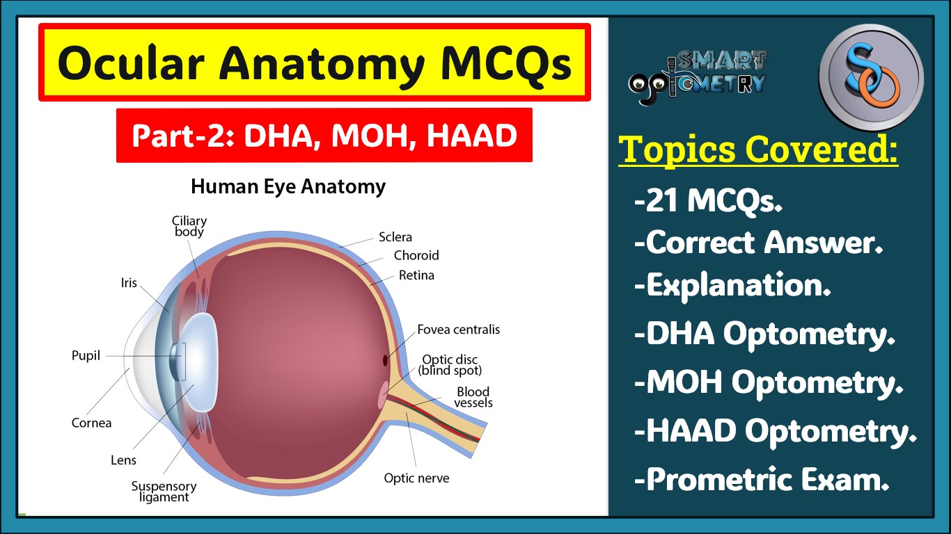
This blog is a must-read for Optometry students and professionals preparing for the Dubai Health Authority (DHA) license exam, HAAD Exam optometry, and Ministry of Health (MOH) license exam. It features 20 meticulously crafted MCQs focused on the Anatomy and Development of the Eye or Ocular Anatomy, covering key topics such as the antero-posterior diameter of the eyeball, the vertical diameter, and the circumference and volume of an adult eyeball.
Each question is provided with detailed answers and explanations, such as the importance of the 24 mm antero-posterior diameter in influencing refractive errors, or how the vertical diameter affects the anatomical structure and function of the eye. You’ll also learn about the anterior and posterior segments of the eyeball, the significance of the crystalline lens’s size and thickness, and the role of Schwalbe’s line in the angle of the anterior chamber.
Whether you’re looking for DHA MCQs online, DHA MCQs for Optometrist, DHA MCQs book, DHA MCQs PDF, DHA exam questions, DHA question paper for optometrists, MOH exam questions, MOH MCQs online, MOH question paper for optometrists, or MOH questions & answer optometry, this blog provides a rich resource that’s essential for acing your exams.
- Check Our Courses: Ophthalmic Instrumentation, Clinical Refraction, Contact Lens, Binocular Vision, Dispensing Optics, MCQs in Optometry
- Download our App “Optometry Notes & MCQs” from Google Play Store.


