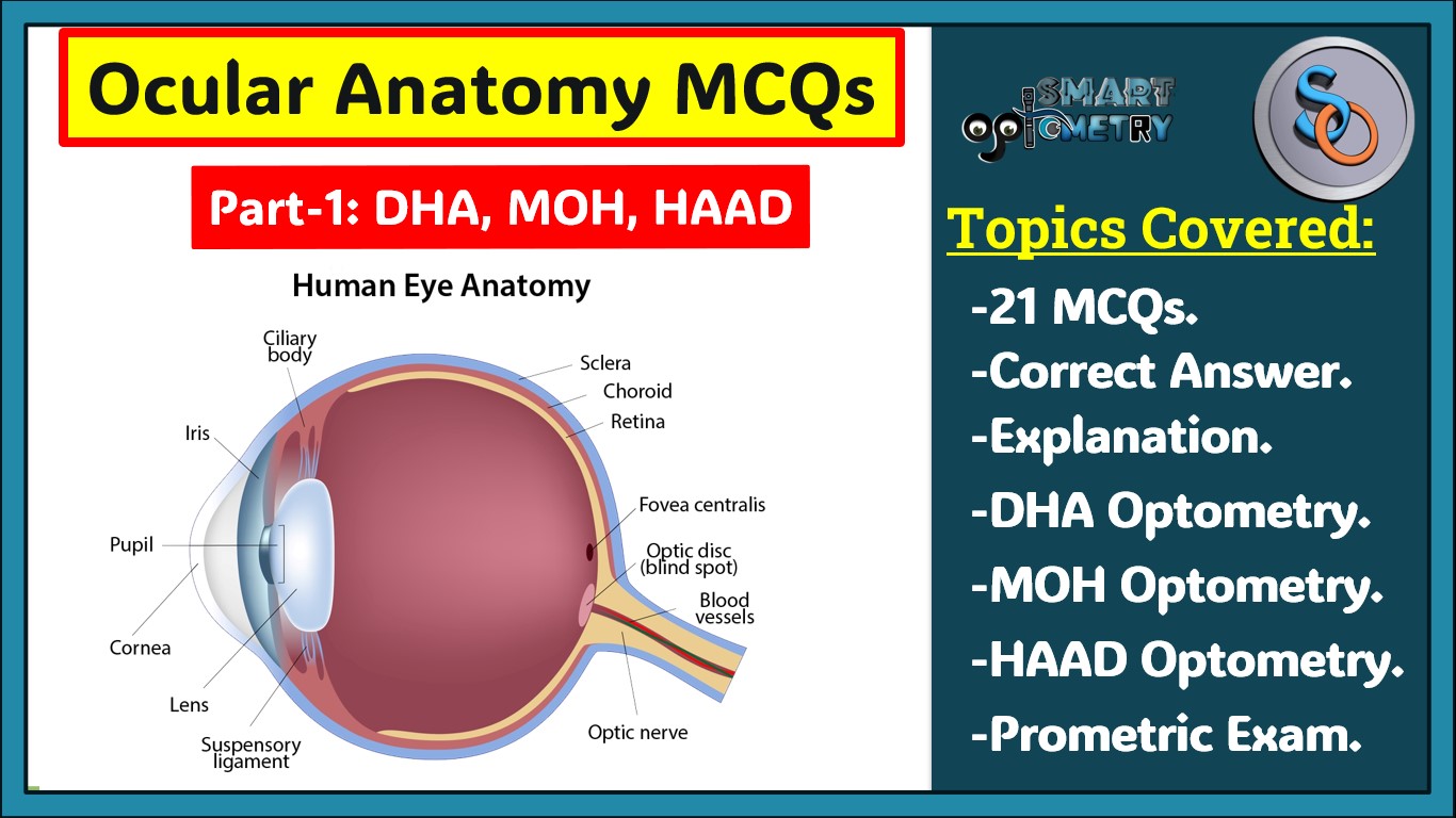
- 1. Antero-posterior diameter of normal adult eyeball is:
- A. 25 mm
- B. 24 mm
- C. 23.5 mm
- D. 23m
Click “Show more” to see the answer and explanation.
Ans: B. 24 mm
The antero-posterior diameter is the longest axis of the eyeball, typically measuring 24 mm in adults. This measurement is essential in determining the axial length, which influences refractive errors. Variations in this diameter can lead to conditions like myopia or hyperopia.
- 2. Smallest diameter of the eyeball is:
- A. Vertical
- B. Horizontal
- C. Antero-posterior
- D. A & B
- 3. Circumference of an adult eyeball is:
- A. 80mm
- B. 65mm
- C. 75mm
- D. 70mm
- 4. Volume of an adult eyeball ls:
- A. 7.5ml
- B. 6.5mL
- C. 5.5mL
- D. 8.0 mL
Click “Show more” to see the answer and explanation.
Ans: B. 6.5mL
The volume of the adult eyeball is around 6.5 mL, which is indicative of the total internal space of the eye. This volume is filled with various fluids like the aqueous and vitreous humor, essential for maintaining intraocular pressure and ocular function.
- 5. Weight of an adult eyeball is:
- A. 7g
- B. 9g
- C. 11 g
- D. 13g
- 6. Anterior segment of the eyeball includes structures lying in front of the:
- A. Iris
- B. Crystalline lens
- C. Vitreous body
- D. Cornea
- 7. Posterior segment of the eyeball includes structures present posterior to the:
- A. Posterior surface of the lens and zonules
- B. Iris and pupil
- C. Vitreous body
- D. Anterior surface of the lens and zonules
- 8. Diameter of an adult crystalline lens is:
- A. 5-6mm
- B. 7-8mm
- C. 9- 10 mm
- D. 11-12mm
- 9. Thickness of the adult crystalline lens is about:
- A. 2.5mm
- B. 3.5mm
- C. 4.00mm
- D. 5mm
- 10. Radius of curvature of the anterior surface of an adult crystalline lens with accommodation at rest is:
- A. 7mm
- B. 10mm
- C. 8mm
- D. 9mm
- 11. Capsule of the crystalline lens is thinnest at:
- A. Anterior pole
- B. Posterior pole
- C. Equator
- D. None of the above
- 12. Infantile nucleus of the crystalline lens refers to the nucleus developed from:
- A. 3 months of gestation to till birth
- B. Birth to one year of age
- C. Birth to puberty
- D. One year of age to 3 years of age
- 13. The lens fibers meet around the Y-shaped suture sin which part of nucleus of the crystalline lens:
- A. Embryonic nucleus
- B. Fetal nucleus
- C. Infantile nucleus
- D. All of the above
Click “Show more” to see the answer and explanation.
Ans: A. Embryonic nucleus
The Y-shaped sutures are characteristic of the embryonic nucleus, which forms early in development. These sutures result from the arrangement of primary lens fibers during the fetal stage. The Y-formation helps in maintaining lens transparency.
- 14. The youngest lens fibers are present in:
- A. Central core of the lens nucleus
- B. Outer layer of the nucleus
- C. Deeper layer of the cortex
- D. Superficial layer of the cortex
Click “Show more” to see the answer and explanation.
Ans: D. Superficial layer of the cortex
The youngest lens fibers are found in the superficial layer of the cortex, as the lens fibers are added from the outside inwards. With age, older fibers are pushed towards the center, forming the nucleus, while new fibers continuously add to the outermost layers.
- 15. Schwalbe’s line forming part of the angle of anterior chamber is the prominent end of:
- A. Sclera
- B. Descemet’s membrane of cornea
- C. Anterior limit of trabecular meshwork
- D. Posterior limit of trabecular meshwork
Click “Show more” to see the answer and explanation.
Ans: B. Descemet’s membrane of cornea
Schwalbe’s line is the termination of Descemet’s membrane at the periphery of the cornea. It is an important landmark in the anatomy of the anterior chamber angle, playing a role in identifying the structures involved in aqueous humor outflow.
- 16. In a normal adult person, the depth of anterior chamber In the centre is about:
- A. 2.5mm
- B. 3mm
- C. 3.5mm
- D. 4mm
Click “Show more” to see the answer and explanation.
Ans: A. 2.5mm
The anterior chamber depth is typically about 2.5 mm in the center of the eye in adults. This depth is crucial for maintaining proper intraocular pressure and for the accommodation of light. Variations in this depth can be associated with conditions like glaucoma.
- 17. ……… is a sweat gland:
- A. Gland of Moll
- B. Gland of Zeis
- C. Meibomian gland
- D. All of the above
- 18. The layer of the cornea once destroyed does not regenerate is:
- A. Epithelium
- B. Bowman’s membrane
- C. Descemet’s membrane
- D. All of the above
Click “Show more” to see the answer and explanation.
Ans: B. Bowman’s membrane
Bowman’s membrane, once destroyed, does not regenerate, which can lead to permanent scarring and vision impairment. It is an acellular layer located beneath the corneal epithelium, providing structural support.
- 19. All of the following are true about corneal Endothelium except:
- A. Cell density is about 3000 cells/mm2 at birth
- B. Corneal decompensation occurs when cell count is decreased by 50%
- C. Endothelial cells contain active pump mechanism
- D. Endothelium is best examined by specular microscopy
- 20. Adult size of the cornea is attained by the age of:
- A. 2 years
- B. 3 years
- C. 5 years
- D. 9 years
Click “Show more” to see the answer and explanation.
Ans: A. 2 years
The cornea reaches its adult size by around 2 years of age, making it one of the earliest parts of the eye to achieve full growth. This rapid growth is essential for normal visual development. After this age, the cornea remains relatively stable in size.
This blog is a must-read for Optometry students and professionals preparing for the Dubai Health Authority (DHA) license exam, HAAD Exam optometry, and Ministry of Health (MOH) license exam. It features 20 meticulously crafted MCQs focused on the Anatomy and Development of the Eye or Ocular Anatomy, covering key topics such as the antero-posterior diameter of the eyeball, the vertical diameter, and the circumference and volume of an adult eyeball.
Each question is provided with detailed answers and explanations, such as the importance of the 24 mm antero-posterior diameter in influencing refractive errors, or how the vertical diameter affects the anatomical structure and function of the eye. You’ll also learn about the anterior and posterior segments of the eyeball, the significance of the crystalline lens’s size and thickness, and the role of Schwalbe’s line in the angle of the anterior chamber. Whether you’re looking for DHA MCQs online, DHA MCQs for Optometrist, DHA MCQs book, DHA MCQs PDF, DHA exam questions, DHA question paper for optometrists, MOH exam questions, MOH MCQs online, MOH question paper for optometrists, or MOH questions & answer optometry, this blog provides a rich resource that’s essential for acing your exams.
- Check Our Courses: Ophthalmic Instrumentation, Clinical Refraction, Contact Lens, Binocular Vision, Dispensing Optics, MCQs in Optometry
- Download our App “Optometry Notes & MCQs” from Google Play Store.
