1) What treatment you will give in unilateral aphakia? (DHA Optometry 2023)
- A. Spectacle.
- B. Contact Lens.
- C. Vision Therapy
- D. Laser Surgery
Click “Show more” to see the answer and explanation.
Ans: B
The most problem with aphakia is high magnification in spectacle lens due to high plus power. In contact lens magnification is only 6-7% where with spectacle magnification is 33%.
Note: Enroll in Course “MCQs in Optometry” in Our App “Optometry Notes & MCQs” to get access to 500+ Previous Year DHA MCQs. 100+ students are preparing and 20+ students already passed DHA entrance.
2) Blood in anterior chamber is known as …….. ? (DHA Optometry 2023)
- A. Hypopyon.
- B. Hyphema.
- C. Hyperopia.
- D. Congestion.
Click “Show more” to see the answer and explanation.
Ans: B
Hyphema is the accumulation of red blood cells (RBCs) in the anterior chamber of the eye.
Note: “MCQs in Optometry” course provide 500+ previous year MCQs, 5000+ subject wise MCQs, 200+ Videos, 100+ Subject wise PDF notes & many more…..
3) What treatment you will give in pediatrics aphakic patient. (DHA Optometry 2023)
- a. IOL followed by Spectacle.
- b. IOL followed by Contact lens.
- c. IOL at the age 10 years.
- d. None of these.
Click “Show more” to see the answer and explanation.
Ans: b
Intraocular lens (IOL) implantation may be considered, especially in older children when the eyes have reached a stable size. However, because the eyes of paediatric patients are still growing, IOLs may not be suitable for very young children. In such cases, contact lenses are often used for optical correction then IOL is planned in later age.
Note: Enroll in Course “MCQs in Optometry” in Our App “Optometry Notes & MCQs” to get access to 500+ Previous Year DHA MCQs. 100+ students are preparing and 20+ students already passed DHA entrance.
4) An increased distance between the orbits, with true lateral displacement of the orbits is called? (DHA Optometry 2022)
- a. Proptosis.
- b. Exophthalmos
- c. Hypertelorism.
- d. None of these.
Click “Show more” to see the answer and explanation.
Ans: c
Orbital hypertelorism is defined as an increased distance between the orbits, with true lateral displacement of the orbits. Anthropometric measurements will yield increased inner canthal distance (ICD), increased outer canthal distance (OCD), and increased interpupillary distance (IPD).
Note: Note: “MCQs in Optometry” course provide 500+ previous year MCQs, 5000+ subject wise MCQs, 200+ Videos, 100+ Subject wise PDF notes & many more…..
5) The interpupillary distance (IPD) is measured between the centers of the pupils is called? (DHA Optometry 2022)
- a. Physiological IPD.
- b. Anatomical IPD.
Click “Show more” to see the answer and explanation.
Ans: b
The interpupillary distance (IPD) is usually measured between the centers of the pupils (anatomical IPD) or between the visual axes (physiological IPD).
Note: Enroll in Course “MCQs in Optometry” in Our App “Optometry Notes & MCQs” to get access to 500+ Previous Year DHA MCQs. 100+ students are preparing and 20+ students already passed DHA entrance.
6) Eikonometer is used to determine? (DHA Optometry 2022)
- a. Aniseikonia.
- b. Polycoria.
- c. Retinal Correspondence.
- d. None of these.
Click “Show more” to see the answer and explanation.
Ans: a
Eikonometer is a device to detect aniseikonia or to test stereoscopic vision
Note: Note: “MCQs in Optometry” course provide 500+ previous year MCQs, 5000+ subject wise MCQs, 200+ Videos, 100+ Subject wise PDF notes & many more…..
7) First clinical sign of Diabetic Retinopathy? (DHA Optometry 2022)
- a. Microaneurysms.
- b. Intraretinal Hemorrhage.
- c. Exudates.
- d. Macular Edema.
Click “Show more” to see the answer and explanation.
Ans: a
The first clinical sign of diabetic retinopathy is often the development of microaneurysms. Microaneurysms are small, localized dilations of small blood vessels in the retina. They can leak fluid and blood into the surrounding retinal tissue, leading to the formation of exudates and the development of other changes characteristic of diabetic retinopathy.
Note: Enroll in Course “MCQs in Optometry” in Our App “Optometry Notes & MCQs” to get access to 500+ Previous Year DHA MCQs. 100+ students are preparing and 20+ students already passed DHA entrance.
8) What is the shape of bony orbit? (DHA Optometry 2024)
- a. Spherical.
- b. Pyramidal
- c. Tubular
- d. None of these.
Click “Show more” to see the answer and explanation.
Ans: b
The orbit of the eye, also known as the eye socket, has a roughly Pyramidal or conical in shape. It is a bony cavity in the skull that houses and protects the eyeball along with its associated structures, such as muscles, nerves, and blood vessels. The orbit is not a perfect cone but has a somewhat irregular shape with various openings and recesses.
Note: Note: “MCQs in Optometry” course provide 500+ previous year MCQs, 5000+ subject wise MCQs, 200+ Videos, 100+ Subject wise PDF notes & many more…..
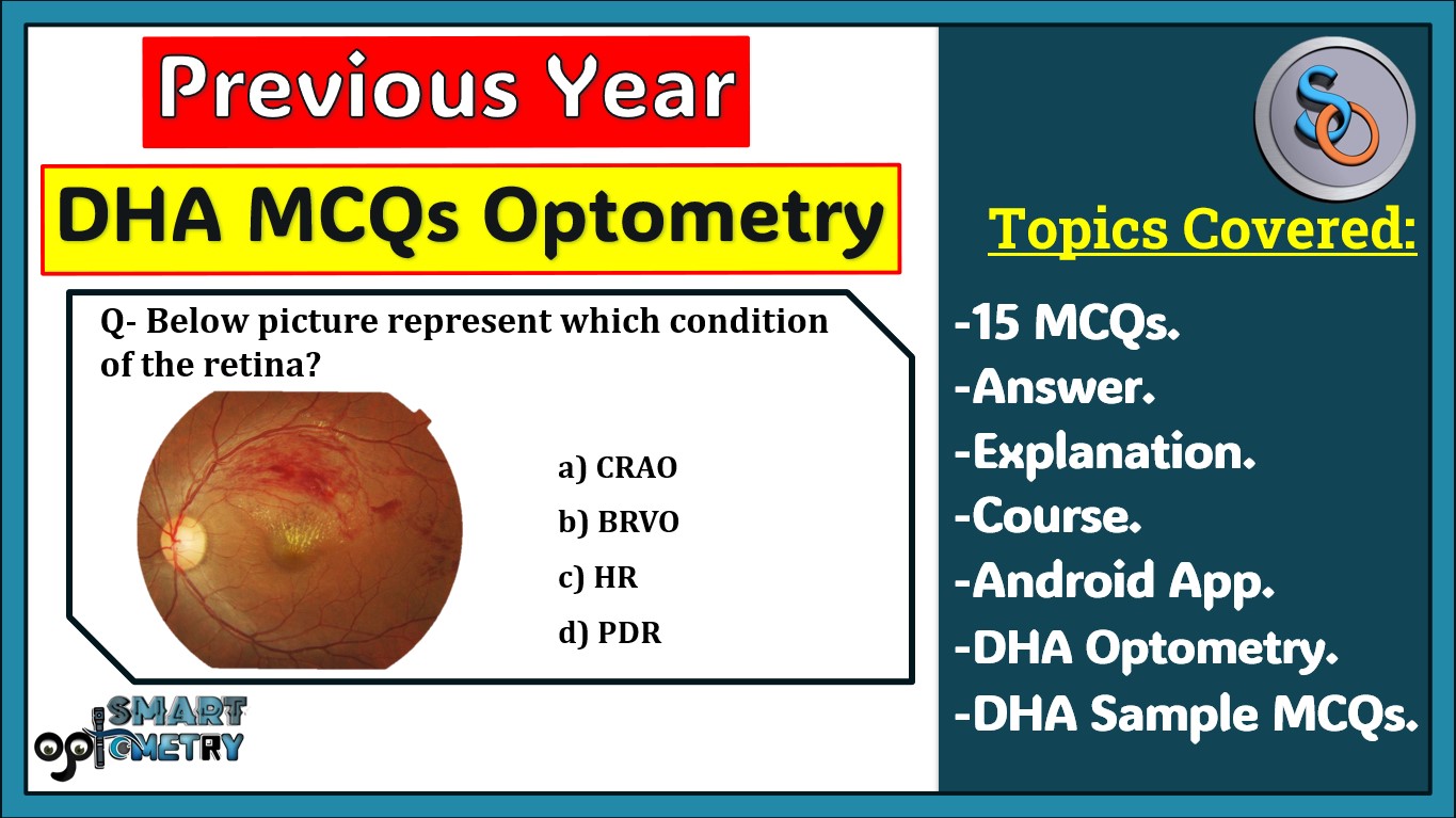
9) Below picture represent which condition of the retina? (DHA Optometry 2024)
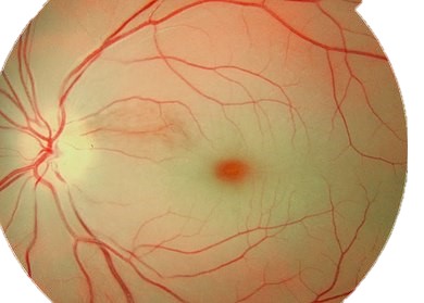
- a) CRAO
- b) BRVO
- c) HR
- d) PDR
Click “Show more” to see the answer and explanation.
Ans: a
In Central Retinal Artery Occlusion, the retina will appear diffusely pale with a cherry red central spot.
Note: Enroll in Course “MCQs in Optometry” in Our App “Optometry Notes & MCQs” to get access to 500+ Previous Year DHA MCQs. 100+ students are preparing and 20+ students already passed DHA entrance.
10) Below picture represent which condition of the retina? (DHA Optometry 2024)
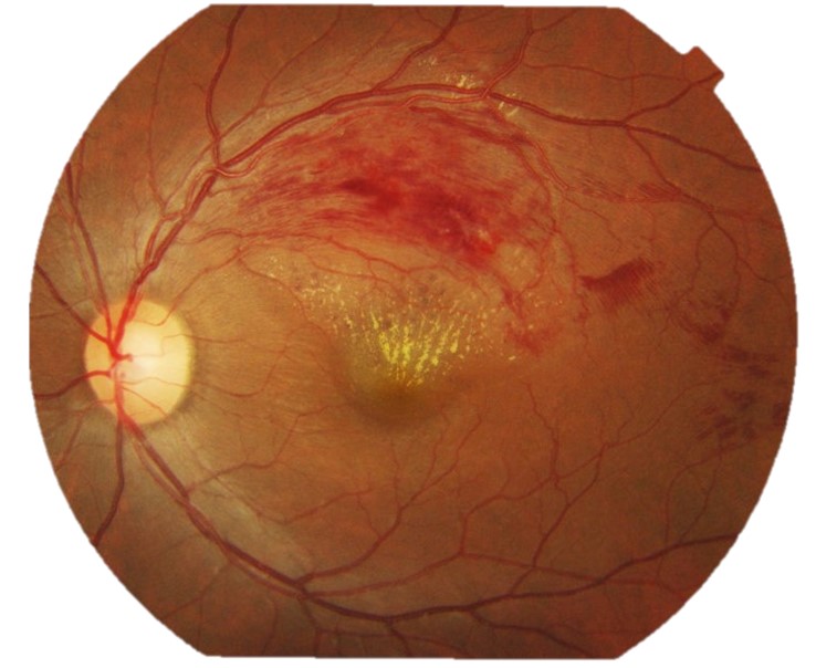
- a) CRAO
- b) BRVO
- c) HR
- d) PDR
Click “Show more” to see the answer and explanation.
Ans: b
Early fundus features of BRVO, include sectorial superficial retinal hemorrhages that rarely cross the horizontal raphe, cotton-wool spots, retinal edema, and dilated and tortuous retinal vein.
Note: Note: “MCQs in Optometry” course provide 500+ previous year MCQs, 5000+ subject wise MCQs, 200+ Videos, 100+ Subject wise PDF notes & many more…..
11) Below picture represent which condition of the retina? (DHA Optometry 2024)
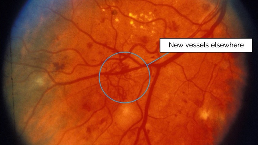
- a) CRAO
- b) BRVO
- c) HR
- d) PDR
Click “Show more” to see the answer and explanation.
Ans: d
Proliferative retinopathy, unlike nonproliferative retinopathy, causes formation of fine preretinal vessel neovascularization visible on the optic nerve or retinal surface. Macular edema or retinal hemorrhage may be visible on funduscopy.
Note: Enroll in Course “MCQs in Optometry” in Our App “Optometry Notes & MCQs” to get access to 500+ Previous Year DHA MCQs. 100+ students are preparing and 20+ students already passed DHA entrance.
12) Metamorphopsia is a visual defect that causes linear objects, such as lines on a grid, to look curvy or rounded. (DHA Optometry 2024)
- a. True
- b. False
Click “Show more” to see the answer and explanation.
Ans: a
Metamorphopsia is a visual defect that causes linear objects, such as lines on a grid, to look curvy or rounded. It’s caused by problems with your eye’s retina, and, in particular, the macula.
Note: Note: “MCQs in Optometry” course provide 500+ previous year MCQs, 5000+ subject wise MCQs, 200+ Videos, 100+ Subject wise PDF notes & many more…..
13) Which type of Cataract is this? (DHA Optometry 2024)
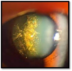
- a. Christmas Tree
- b. Sunflower cataracts
- c. Snowflake type cataract
- d. None of these
Click “Show more” to see the answer and explanation.
Ans: a
It consists of small white dots arranged in a pattern resembling a Christmas tree, hence its name. The dots are semi-transparent and can form a cloudy haze within the lens that obstructs vision. This condition may affect one or both eyes, and it can lead to a loss of vision if left untreated.
Note: Enroll in Course “MCQs in Optometry” in Our App “Optometry Notes & MCQs” to get access to 500+ Previous Year DHA MCQs. 100+ students are preparing and 20+ students already passed DHA entrance.
14) What is the outermost layer of choroid? (DHA Optometry 2024)
- a. Lamina Fusca
- b. Basal Lamina
- c. Choriocapillaris
- d. Stroma of Choroid
Click “Show more” to see the answer and explanation.
Ans: a
It is a thin (10-34 µm) membrane of condensed collagen fibres, melanocytes and fibroblasts. It is continuous anteriorly with the supraciliary lamina of the ciliary body. The potential space between this membrane and sclera is called suprachoroidal space which contains long and short posterior ciliary arteries and nerves.
Note: Note: “MCQs in Optometry” course provide 500+ previous year MCQs, 5000+ subject wise MCQs, 200+ Videos, 100+ Subject wise PDF notes & many more…..
15) Which cells are affected in Retinitis pigmentosa? (DHA Optometry 2024)
- a. Photoreceptor Cells.
- b. Bipolar Cells.
- c. Amacrine Cells.
- d. None of these.
Click “Show more” to see the answer and explanation.
Ans: a
Retinitis Pigmentosa (RP) is a group of diseases in which one of a large number of mutations causes death of rod photoreceptors. After rods die, cone photoreceptors slowly degenerate in a characteristic pattern.
Note: Enroll in Course “MCQs in Optometry” in Our App “Optometry Notes & MCQs” to get access to 500+ Previous Year DHA MCQs. 100+ students are preparing and 20+ students already passed DHA entrance.
- Check Our Courses: Ophthalmic Instrumentation, Clinical Refraction, Contact Lens, Binocular Vision, Dispensing Optics, MCQs in Optometry
- Download our App “Optometry Notes & MCQs


5 Comments
Know Everything about DHA: https://smartoptometryacademy.com/dha-exam-for-optometrists/
I want to learn about DHA
helloI really like your writing so a lot share we keep up a correspondence extra approximately your post on AOL I need an expert in this house to unravel my problem May be that is you Taking a look ahead to see you
Stay with us and share with your friends for more…………..
Thank you