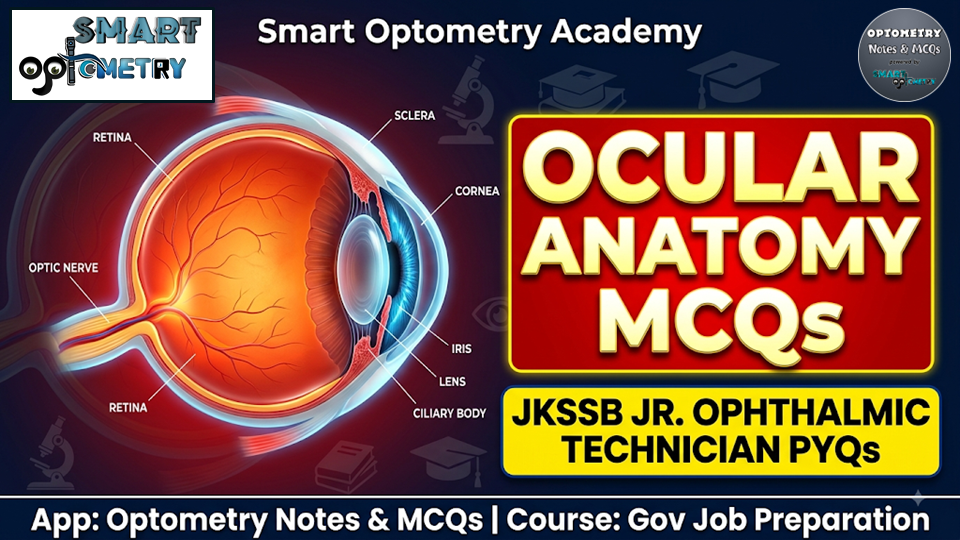
Master Ocular Anatomy with JKSSB Previous Year Questions
Ocular Anatomy forms the bedrock of the written examination for technical eye care vacancies under the Jammu & Kashmir Services Selection Board (JKSSB). Whether you are targeting the Health and Medical Education Department’s competitive examinations or solidifying your foundational knowledge, practicing authentic JKSSB Ophthalmic Technician Previous Year MCQs is the most effective way to understand the exam pattern, question framing, and high-yield topics.
This curated question bank focuses exclusively on core anatomical structures, embryological development, extraocular muscle actions, and visual pathways. Reviewing these JKSSB Ophthalmic Technician PYQs will help you avoid the critical 0.25 negative marking penalty and build the clinical reasoning required for a top merit rank.
Why Ocular Anatomy is High-Yield in JKSSB Examinations
In state-level paramedical and ophthalmic officer tests, anatomy questions evaluate both microscopic retinal architecture and macroscopic orbital landmarks. By solving these JKSSB Optometry PYQs, candidates can review essential high-frequency themes, including:
- Retinal Architecture & Photoreceptors: Understanding why the fovea centralis lacks rods (leading to physiological foveal night-blindness) and how cone photopigments facilitate photopic vision and color discrimination.
- Anterior Segment & Fluid Dynamics: Differentiating the iris’s pupillary and ciliary zones divided by the collarette, and tracing the trabecular outflow of aqueous humor through the Canal of Schlemm.
- Orbital Topography & Extraocular Muscles: Pinpointing the common tendinous origin (Annulus of Zinn) of all four rectus muscles at the orbital apex and mastering primary vs. secondary muscle kinematics (such as the intorsion action of the superior rectus).
- Neuro-Ophthalmology & Vasculature: Mapping the visual pathway from retinal ganglion cell axons forming the optic nerve, through nasal fiber decussation at the optic chiasma, to orbital venous drainage via the cavernous sinus.
- Ocular Embryology & Development: Key developmental milestones, such as neonatal eye dimensions (the eyeball is roughly 70% of adult volume at birth) and the role of the primitive hyaloid artery in forming the tunica vasculosa lentis.
Practice BFUHS Previous Year MCQs on “Pediatric Optometry”:
Q-1: The normal fovea is “blind” in dim light conditions. This phenomenon is known as normal foveal night-blindness and can be explained by which of the following? (JKSSB Ophthalmic Technician Previous Year MCQs)
A) Absence of rods in the fovea
B) Absence of cones in the fovea
C) Thickening of the retinal pigment epithelium
D) Lack of blood supply to the fovea
Struggling with your optometry government job preparation? Let Samir Sir, the founder of Smart Optometry, guide you to success. Enroll in our “Gov Job Preparation” course, and we’ll provide the roadmap. Your dream job at BFUHS Ophthalmic Officer, GSSSB, AIIMS, RRB, or DHS is within reach!
Q-2: Which of the following options is the part of the Iris, that lies between the Collarette and Pigmented frill?(JKSSB Ophthalmic Technician PYQs)
A) Pupillary zone
B) Contraction furrow
C) Ciliary zone
D) Crypts
Click “Show more” to see the answer and explanation.
nswer: A) Pupillary zone
The collarette divides the iris into an inner pupillary zone extending to the pigmented pupillary frill, and an outer ciliary zone toward the iris root. Contraction furrows and crypts are surface features within these zones.
.
.
Q-3: The ciliary body is responsible for the production of aqueous humor. The aqueous humor drains primarily through: (JKSSB Ophthalmic Technician Previous Year MCQs)
A) The vortex veins
B) The canal of Schlemm
C) The central retinal vein
D) The suprachoroidal space
Unlock your potential with our “Gov Job Preparation” course. Gain access to a massive library of 5000+ subject-wise MCQs, 1000+ previous year questions, detailed notes, and video lectures. Everything you need for exams like BFUHS Ophthalmic Officer, AIIMS, GSSSB, PSC, and DMER is in one place.
Q-4: The optic nerve (CN II) carries visual information from the retina to the brain. It is formed by the axons of: (JKSSB Ophthalmic Technician PYQs)
A) Bipolar cells
B) Ganglion cells
C) Photoreceptor cells
D) Amacrine cells
.
.
Q-5: The central retinal artery is a branch of the ophthalmic artery. It enters the optic nerve at the: (JKSSB Ophthalmic Technician Previous Year MCQs)
A) Optic chiasm
B) Orbital apex
C) Intracanalicular portion of the optic nerve
D) Lamina cribrosa
Click “Show more” to see the answer and explanation.
Answer: B) Orbital apex
The central retinal artery pierces the dural sheath and enters the substance of the optic nerve within its intraorbital portion, precisely 10 to 12 mm behind the globe. It then travels centrally inside the core of the nerve, eventually passing through the lamina cribrosa at the optic disc to supply the inner layers of the retina.
Prepare for a full year of opportunities! Our “Gov Job Preparation” course gives you 12 months of access to top-quality materials and support. Cover all major exams like BFUHS Ophthalmic Officer, GSSSB, AIIMS, RRB, Ophthalmic Officer without buying multiple courses. It’s the smartest investment for your career.
Q-6: Which is the area in visual pathway, the nerve fibres from nasal halves of the two retinae decussate? (JKSSB Ophthalmic Technician PYQs)
A) Optic tract
B) Optic nerve
C) Optic radiation
D) Optic chiasma
Click “Show more” to see the answer and explanation.
Answer: D) Optic chiasma
At the optic chiasm, axons from the nasal retina of each eye cross to the opposite side, while temporal fibers remain uncrossed. This decussation is fundamental for binocular visual field representation.
.
.
Q-7: Venous drainage of the orbit occurs primarily through the superior and inferior ophthalmic veins, which drain into the: (JKSSB Ophthalmic Technician Previous Year MCQs)
A) Cavernous sinus
B) Internal jugular vein
C) Pterygoid plexus
D) External jugular vein
Don’t study alone! Our “Gov Job Preparation” course includes group updates and live support to clear all your doubts. With guidance from Samir Sir and a community of fellow aspirants, you’ll stay motivated and on track.
Q-8: All the four rectus muscles of the eye originate from? (JKSSB Ophthalmic Technician PYQs)
A) Orbital roof
B) Lateral wall
C) Orbital floor
D) Annulus of Zinn
Click “Show more” to see the answer and explanation.
Answer: D) Annulus of Zinn
The superior, inferior, medial, and lateral rectus muscles all arise from the common tendinous ring (annulus of Zinn) at the orbital apex, which also encircles the optic foramen and superior orbital fissure.
.
.
Q-9: Which photoreceptors has the function of Color Vision? (JKSSB Ophthalmic Technician Previous Year MCQs)
A) Amacrine cells
B) Cones
C) Rods
D) Bipolar cells
Ready to secure your government job in optometry? The “Gov Job Preparation” course is your final destination. With a proven success strategy, expert mentorship, and over 6000 MCQs, we make your preparation seamless and effective. Click the link to begin your journey!
.
Q-10: What is the secondary action of the superior rectus muscle? (JKSSB Ophthalmic Technician PYQs)
A) Elevation
B) Intorsion
C) Abduction
D) Extortion
.
🔒 Bonus MCQs – Available in App
Unlock these 10 exclusive premium questions + detailed explanations in the Govt Job Preparation course.
Q-11: Which of these help in visualization of corneal cells?
A Confocal microscope
B Optical coherence tomography
C Pachymeter
D Slit lamp biomicroscope
Answer: A) Confocal microscope
A confocal microscope allows detailed, in vivo imaging of corneal cells, including endothelial and epithelial layers. It provides high-resolution cellular-level imaging essential for diagnosing corneal pathologies.
Q-12: Which are not the uses of single or double frequency Nd:YAG laser?
A Spectacle removal
B Laser iridotomy
C Pan retinal photocoagulation in diabetic retinopathy
D Ablation of posterior capsular opacification
Answer: A) Spectacle removal
Nd:YAG lasers are used for iridotomy, posterior capsular opacification ablation, and sometimes diabetic retinopathy treatment. They cannot be used for spectacle removal, which requires refractive surgery like LASIK.
Q-13: Slit lamp biomicroscope cannot be used for which of the following:
A Measurement of corneal epithelial defect
B Observing the optic disc and macula with 90D lens
C Laser iridectomy in angle closure glaucoma
D Fluorescein staining with cobalt blue light
Answer: C) Laser iridectomy in angle closure glaucoma
A slit lamp is a diagnostic tool and cannot perform surgical procedures like laser iridectomy. Such procedures require specific laser devices designed for therapeutic interventions.
Q-14: Gold standard for measurement of IOP is:
A Digital tonometry
B Schiotz tonometry
C Pneumotonometry
D Applanation tonometry
Answer: D) Applanation tonometry
Goldmann Applanation Tonometry is widely regarded as the gold standard for measuring intraocular pressure. It works by measuring the force required to flatten a fixed area of the cornea and is considered the most accurate method for glaucoma diagnosis and management.
Q-15: Which laser is used in the management of after-cataract?
A Argon
B Krypton
C Nd:YAG
D Excimer
Answer: C) Nd:YAG
The Nd:YAG laser is the standard tool used for posterior capsulotomy to disrupt opacified posterior capsule. This restores clear vision in patients who develop after-cataract following cataract surgery.
Q-16: Instrument used to measure phoria for near (33 cm) is:
A Maddox rod
B Maddox wing
C RAF rule
D None of the above
Answer: B) Maddox wing
The Maddox wing is specifically designed for measuring heterophoria at near distance (typically 33 cm). It uses a combination of red and white lights with a dissociating effect to quantify the deviation.
Q-17: Auto-refractometer is a method of refraction:
A Subjective
B Objective
C Both of the above
D None of the above
Answer: B) Objective
Auto-refractometry is an objective method of refraction that measures the eye’s refractive error without requiring patient responses. It provides a reliable starting point for subjective refraction.
Q-18: Jackson’s cross cylinder is used to check:
A Axis of the cylinder
B Power of the cylinder
C Both of the above
D None of the above
Answer: C) Both of the above
The Jackson Cross Cylinder is used to refine both the axis and power of the cylindrical correction during subjective refraction. It allows precise adjustment of astigmatic correction by presenting two alternative lens positions.
Q-19: Keratometry measures:
A Retinal thickness
B Corneal curvature
C Lens power
D Visual fields
Answer: B) Corneal curvature
Keratometry measures the radius of curvature of the anterior corneal surface. This measurement is essential for contact lens fitting, IOL power calculation, and diagnosing corneal conditions like keratoconus.
Q-20: Which instrument is used to measure intraocular pressure?
A Perimeter
B Retinoscope
C Tonometer
D Ophthalmoscope
Answer: C) Tonometer
A tonometer is the instrument used to measure intraocular pressure, with Goldmann Applanation Tonometry being the gold standard. Regular IOP measurement is essential for glaucoma diagnosis and management.
🎯 Accelerate Your Optometry Exam Prep
Access high-yield study resources, smart mock tests, and a high-performance community.
🎯 Enroll in Full Govt Job Preparation Course.
JKSSB Ophthalmic Technician 2026
Previous Year Questions (PYQs)
.
Master Your Gov Job Prep
Join our comprehensive “Gov Job Preparation” course available exclusively on the Optometry Notes & MCQs App.
The Ultimate Success Strategy
Follow this proven sequence to maximize your preparation and secure your goal.
Subjects wise Theory Notes
Build a strong foundation by mastering the core theoretical concepts for each subject.
Premium Lectures & Videos
Deepen your understanding with expert video explanations and fundamental optometry lectures.
Subjects wise MCQs
Test your knowledge and practice applying concepts with targeted multiple-choice questions.
Previous Year MCQs
Familiarize yourself with actual exam patterns and gauge your readiness with past papers.
Ready to Start Your Journey?
.
About the JKSSB Junior Ophthalmic Technician Recruitment
The JKSSB Junior Ophthalmic Technician recruitment conducted by the Jammu & Kashmir Services Selection Board is one of the most sought-after opportunities for eye care professionals seeking government positions under the Health and Medical Education Department. Released periodically to fulfill technical healthcare requirements across various district and divisional cadres, this JKSSB optometry vacancy offers a Level-4 pay scale position dedicated to primary clinical assistance, vision screening, and ophthalmic diagnostic support.
Securing a post through the JKSSB Ophthalmic Technician Recruitment requires thorough preparation across specialized ophthalmic subjects as well as general medical sciences. Because selection is determined purely on merit scored in the written objective examination, mastering subject-wise Multiple Choice Questions (MCQs) and understanding the official exam pattern are essential steps toward qualifying.
Key Highlights of the JKSSB Optometry Vacancy
- Recruiting Body: Jammu & Kashmir Services Selection Board (JKSSB)
- Department: Health and Medical Education Department
- Post Title: Junior Ophthalmic Technician / Ophthalmic Assistant
- Pay Scale: Level-4
- Cadres: Divisional and District Cadres (UT of Jammu & Kashmir)
- Selection Mode: Objective Type Written Examination (OMR / Computer-Based Test)
- Language Medium: English Only
Prescribed Educational Qualification & Eligibility
To apply under the JKSSB Junior Ophthalmic Technician recruitment, candidates must satisfy specific educational benchmarks set by the recruitment board:
- Basic Qualification: Matriculation (10th standard) or above from a recognized board.
- Technical Qualification: A recognized Degree or Diploma in Ophthalmic Technician Course / Ophthalmic Techniques from a state- or central-recognized medical institute or paramedical council.
- Equivalence for Optometry Graduates: Candidates holding higher qualifications such as a Bachelor of Science (B.Sc.) in Optometry or a Diploma in Optometry must ensure their qualification is backed by an official Equivalence Certificate from the competent authority/university if required during document verification.
- Domicile Criteria: Applicants must hold a valid Domicile Certificate of the Union Territory of Jammu & Kashmir.
Examination Pattern & Marking Scheme
The selection process for the JKSSB Ophthalmic Technician Recruitment involves a competitive written test structured as follows:
- Question Format: Multiple Choice Questions (MCQs) covering foundational and advanced ophthalmic concepts.
- Negative Marking: There is a penalty of 0.25 (one-fourth) marks for every incorrect answer.
- Core Technical Focus: The majority of the syllabus is dedicated to domain-specific subjects, including Ocular Anatomy, Ocular Physiology, Optics, Refraction, Ophthalmic Instrumentation, Ocular Pathology, Pharmacology, and Community Ophthalmology.
- Merit-Based Allocation: Final appointment lists are drafted strictly on the basis of written test scores combined with candidate cadre preferences and successful document verification.
Why Subject-Wise MCQ Practice is Crucial for Selection
Success in the JKSSB Junior Ophthalmic Technician recruitment examination depends on high accuracy and rapid recall under time pressure. Reviewing past questions by individual subject helps candidates:
- Pinpoint high-yield topics frequently targeted by the JKSSB exam panel.
- Master difficult optical formulas, anatomical landmarks, and pharmacological contraindications.
- Minimize negative marking by testing diagnostic reasoning before the actual test.
- Bridge the conceptual gap between theoretical optometry textbooks and clinical technician-level applications.
Use the curated previous year questions and topic-wise mock tests provided in this series to build subject mastery and secure your seat in the JKSSB optometry vacancy.
👉 View Full Course Overview & Success Stories
📲 Download the Optometry Notes & MCQs App on Google Play Store