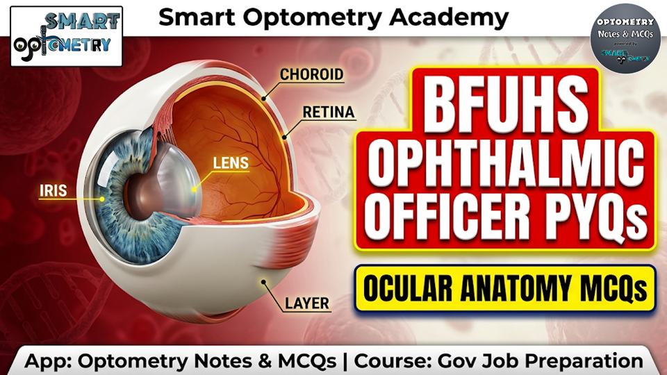
BFUHS Ophthalmic Officer Previous Year MCQs: Ocular Anatomy Solved Paper
Prepare for your upcoming state optometry recruitment exams with our high-yield collection of BFUHS Ophthalmic Officer Previous Year MCQs focused on the essential subject of Ocular Anatomy. To secure a top rank in the written examination, consistent practice with authentic Ophthalmic Officer MCQs is absolutely critical. This blog provides a carefully curated set of Ophthalmic Officer Previous Year MCQs designed to strengthen your anatomical foundations, improve your accuracy, and boost your overall exam score.
Ocular Anatomy is a core subject that tests your in-depth understanding of the eye’s intricate structural framework, nerve pathways, and muscular dynamics. This practice module breaks down highly tested questions with detailed clinical explanations to ensure maximum retention. In this comprehensive practice set, you will review and master the following key anatomical concepts:
- Anterior Segment & Uveal Tract: Identifying key structural landmarks like Schwalbe’s line (the termination of Descemet’s membrane), understanding the continuous formation of new lens fibres in the superficial cortex, and distinguishing components of the uveal tract.
- Extraocular & Intraocular Muscles: Mastering the primary, secondary, and tertiary actions of extraocular muscles (such as extorsion by the inferior rectus and intorsion/depression by the superior oblique) and identifying intrinsic smooth muscles like the ciliary muscle.
- Cranial Nerve Innervations: Reviewing critical nerve supplies, including the innervation of the inferior oblique by the 3rd cranial nerve (Oculomotor nerve) and the classification of the optic nerve as the 2nd cranial nerve.
- Glandular Functions & Ocular Movements: Understanding the secretion of the tear film’s lipid layer by the Meibomian glands, the primary role of the Levator Palpebrae Superioris in eyelid elevation, and differentiating monocular movements (abduction) from binocular versions.
Practice BFUHS Previous Year MCQs on “Ocular Anatomy”:
Q-1: Schwalbe’s line forming part of the angle of the anterior chamber is the prominent end of: (BFUHS Ophthalmic Officer Previous Year MCQs)
A) Sclera
B) Descemet’s membrane of cornea
C) Anterior limit of trabecular meshwork
D) Posterior limit of trabecular meshwork
Struggling with your optometry government job preparation? Let Samir Sir, the founder of Smart Optometry, guide you to success. Enroll in our “Gov Job Preparation” course, and we’ll provide the roadmap. Your dream job at BFUHS Ophthalmic Officer, GSSSB, AIIMS, RRB, or DHS is within reach!
Q-2: The youngest lens fibres are present in: (BFUHS Optometry Previous Year Question Paper)
A) Central core of the lens nucleus
B) Outer layer of the nucleus
C) Deeper layer of the cortex
D) Superficial layer of the cortex
Click “Show more” to see the answer and explanation.
Answer: D) Superficial layer of the cortex
The most recently formed lens fibres are located in the superficial cortex. New lens fibres are continuously added throughout life, forming the characteristic layered structure of the lens.
.
.
Q-3: Anterior segment of the eyeball includes structures lying in front of the: (BFUHS Ophthalmic Officer Previous Year MCQs)
A) Iris
B) Crystalline lens
C) Vitreous body
D) Cornea
Click “Show more” to see the answer and explanation.
Answer: C) Vitreous body
The anterior segment includes structures in front of the vitreous body: cornea, anterior and posterior chambers, iris, ciliary body, and crystalline lens. The vitreous body lies in the posterior segment.
Unlock your potential with our “Gov Job Preparation” course. Gain access to a massive library of 5000+ subject-wise MCQs, 1000+ previous year questions, detailed notes, and video lectures. Everything you need for exams like BFUHS Ophthalmic Officer, AIIMS, GSSSB, PSC, and DMER is in one place.
Q-4: All of the following are a part of uvea EXCEPT:(BFUHS Optometry Previous Year Question Paper)
A) Iris
B) Ciliary body
C) Choroid
D) Schwalbe’s line
Click “Show more” to see the answer and explanation.
Answer: D) Schwalbe’s line
The uveal tract consists of three parts: the iris, the ciliary body, and the choroid. Schwalbe’s line is an anatomical landmark in the anterior chamber angle representing the termination of Descemet’s membrane, not a uveal structure.
.
.
Q-5: The lipid layer of Tear film is produced by: (BFUHS Ophthalmic Officer Previous Year MCQs)
A) Meibomian glands
B) Main lacrimal gland
C) Accessory lacrimal glands
D) Goblet cells
Click “Show more” to see the answer and explanation.
Answer: A) Meibomian glands
The tear film’s outermost lipid layer is secreted primarily by the Meibomian glands located in the tarsal plates. This oily layer prevents rapid evaporation of the underlying aqueous layer and stabilizes the tear film.
Prepare for a full year of opportunities! Our “Gov Job Preparation” course gives you 12 months of access to top-quality materials and support. Cover all major exams like BFUHS Ophthalmic Officer, GSSSB, AIIMS, RRB, Ophthalmic Officer without buying multiple courses. It’s the smartest investment for your career.
Q-6: The main elevator of the eyelids is:(BFUHS Ophthalmic officer Previous Year Question Paper)
A) Levator palpebrae superioris
B) Muller’s muscle
C) Inferior rectus
D) Obicularis oculi
Click “Show more” to see the answer and explanation.
Answer: A) Levator palpebrae superioris
The Levator Palpebrae Superioris is the primary muscle responsible for lifting the upper eyelid. Muller’s muscle provides a small amount of accessory elevation, but the LPS is the main functional elevator.
.
.
Q-7: The optic nerve, also known as:(BFUHS Ophthalmic Officer Previous Year MCQs)
A) First cranial nerve
B) Second cranial nerve
C) Third cranial nerve
D) Fourth cranial nerve
Click “Show more” to see the answer and explanation.
Answer: B) Second cranial nerve
The optic nerve is anatomically classified as the second cranial nerve (CN II), responsible for transmitting visual information from the retina to the brain. It is unique because it acts more like a tract of the central nervous system than a peripheral nerve.
Don’t study alone! Our “Gov Job Preparation” course includes group updates and live support to clear all your doubts. With guidance from Samir Sir and a community of fellow aspirants, you’ll stay motivated and on track.
Q-8: Movement caused by superior oblique muscle: (BFUHS Ophthalmic officer Previous Year Question Paper)
A) Depression
B) Intorsion
C) None of the above
D) Both a and b
.
.
Q-9: Nerve supply of inferior oblique muscle is by: (BFUHS Ophthalmic Officer Previous Year MCQs)
A) 3rd Cranial Nerve
B) 4th Cranial Nerve
C) 6th Cranial Nerve
D) 7th Cranial Nerve
Ready to secure your government job in optometry? The “Gov Job Preparation” course is your final destination. With a proven success strategy, expert mentorship, and over 6000 MCQs, we make your preparation seamless and effective. Click the link to begin your journey!
.
Q-10: What is the secondary action of the Inferior Rectus Muscle? (BFUHS Ophthalmic Officer Previous Year MCQs)
A) Adduction
B) Extorsion
C) Depression
D) None of these
Click “Show more” to see the answer and explanation.
Answer: B) Extorsion
The primary action of the inferior rectus is depression, its secondary action is extorsion (excycloduction), and its tertiary action is adduction. This is because the muscle plane forms a 23-degree angle with the visual axis.
.
.
Q-11: Which of the following is the example of Intra-ocular muscle?(BFUHS Ophthalmic Officer Previous Year MCQs)
A) Levator palpebrae superioris
B) Ciliary muscle
C) Inferior tarsal muscle
D) Medial rectus
Unlock your potential with our “Gov Job Preparation” course. Gain access to a massive library of 5000+ subject-wise MCQs, 1000+ previous year questions, detailed notes, and video lectures. Everything you need for exams like BFUHS Ophthalmic Officer, AIIMS, GSSSB, PSC, and DMER is in one place.
Q-12: Which of the examples of the monocular movement? (BFUHS Optometry Previous Year Question Paper)
A) Levoversion
B) Dextroversion
C) Divergence
D) Abduction
Click “Show more” to see the answer and explanation.
Answer: D) Abduction
Abduction is a duction referring to the movement of a single eye rotating away from the midline. Levoversion and dextroversion are versions (conjugate binocular movements), and divergence is a vergence (disjunctive binocular movement).
🔒 Bonus MCQs – Available in App
Unlock these 10 exclusive premium questions + detailed explanations in the Govt Job Preparation course.
Q-11: Which of these help in visualization of corneal cells?
A Confocal microscope
B Optical coherence tomography
C Pachymeter
D Slit lamp biomicroscope
Answer: A) Confocal microscope
A confocal microscope allows detailed, in vivo imaging of corneal cells, including endothelial and epithelial layers. It provides high-resolution cellular-level imaging essential for diagnosing corneal pathologies.
Q-12: Which are not the uses of single or double frequency Nd:YAG laser?
A Spectacle removal
B Laser iridotomy
C Pan retinal photocoagulation in diabetic retinopathy
D Ablation of posterior capsular opacification
Answer: A) Spectacle removal
Nd:YAG lasers are used for iridotomy, posterior capsular opacification ablation, and sometimes diabetic retinopathy treatment. They cannot be used for spectacle removal, which requires refractive surgery like LASIK.
Q-13: Slit lamp biomicroscope cannot be used for which of the following:
A Measurement of corneal epithelial defect
B Observing the optic disc and macula with 90D lens
C Laser iridectomy in angle closure glaucoma
D Fluorescein staining with cobalt blue light
Answer: C) Laser iridectomy in angle closure glaucoma
A slit lamp is a diagnostic tool and cannot perform surgical procedures like laser iridectomy. Such procedures require specific laser devices designed for therapeutic interventions.
Q-14: Gold standard for measurement of IOP is:
A Digital tonometry
B Schiotz tonometry
C Pneumotonometry
D Applanation tonometry
Answer: D) Applanation tonometry
Goldmann Applanation Tonometry is widely regarded as the gold standard for measuring intraocular pressure. It works by measuring the force required to flatten a fixed area of the cornea and is considered the most accurate method for glaucoma diagnosis and management.
Q-15: Which laser is used in the management of after-cataract?
A Argon
B Krypton
C Nd:YAG
D Excimer
Answer: C) Nd:YAG
The Nd:YAG laser is the standard tool used for posterior capsulotomy to disrupt opacified posterior capsule. This restores clear vision in patients who develop after-cataract following cataract surgery.
Q-16: Instrument used to measure phoria for near (33 cm) is:
A Maddox rod
B Maddox wing
C RAF rule
D None of the above
Answer: B) Maddox wing
The Maddox wing is specifically designed for measuring heterophoria at near distance (typically 33 cm). It uses a combination of red and white lights with a dissociating effect to quantify the deviation.
Q-17: Auto-refractometer is a method of refraction:
A Subjective
B Objective
C Both of the above
D None of the above
Answer: B) Objective
Auto-refractometry is an objective method of refraction that measures the eye’s refractive error without requiring patient responses. It provides a reliable starting point for subjective refraction.
Q-18: Jackson’s cross cylinder is used to check:
A Axis of the cylinder
B Power of the cylinder
C Both of the above
D None of the above
Answer: C) Both of the above
The Jackson Cross Cylinder is used to refine both the axis and power of the cylindrical correction during subjective refraction. It allows precise adjustment of astigmatic correction by presenting two alternative lens positions.
Q-19: Keratometry measures:
A Retinal thickness
B Corneal curvature
C Lens power
D Visual fields
Answer: B) Corneal curvature
Keratometry measures the radius of curvature of the anterior corneal surface. This measurement is essential for contact lens fitting, IOL power calculation, and diagnosing corneal conditions like keratoconus.
Q-20: Which instrument is used to measure intraocular pressure?
A Perimeter
B Retinoscope
C Tonometer
D Ophthalmoscope
Answer: C) Tonometer
A tonometer is the instrument used to measure intraocular pressure, with Goldmann Applanation Tonometry being the gold standard. Regular IOP measurement is essential for glaucoma diagnosis and management.
🎯 Accelerate Your Optometry Exam Prep
Access high-yield study resources, smart mock tests, and a high-performance community.
🎯 Enroll in Full Govt Job Preparation Course.
BFUHS Ophthalmic Officer 2026
Previous Year Questions (PYQs)
.
Master Your Gov Job Prep
Join our comprehensive “Gov Job Preparation” course available exclusively on the Optometry Notes & MCQs App.
The Ultimate Success Strategy
Follow this proven sequence to maximize your preparation and secure your goal.
Subjects wise Theory Notes
Build a strong foundation by mastering the core theoretical concepts for each subject.
Premium Lectures & Videos
Deepen your understanding with expert video explanations and fundamental optometry lectures.
Subjects wise MCQs
Test your knowledge and practice applying concepts with targeted multiple-choice questions.
Previous Year MCQs
Familiarize yourself with actual exam patterns and gauge your readiness with past papers.
Ready to Start Your Journey?
📋 Key Recruitment Highlights & Exam Pattern:
- Recruiting Body: Baba Farid University of Health Sciences (BFUHS), Faridkot (on behalf of the Directorate of Health & Family Welfare, Punjab).
- Pay Scale: Initial Pay Scale is ₹10,300–34,800 + Grade Pay ₹3,800, with a revised 7th Pay Commission convergence starting salary of ₹35,400/- per month.
- Essential Qualification: Must have passed 10+2 in Science with Biology (or equivalent) AND hold a Diploma in Ophthalmic Assistant from a recognized university or institution. (Note: As per Term & Condition No. 20, candidates with higher qualifications like BSc Optometry can apply, but no extra weightage or bonus marks will be given for higher qualifications).
- Selection Process & Weightage (100 Marks Total):
- Written Examination: 90 Marks (Objective MCQ test conducted by BFUHS). Candidates must score a minimum of 33% marks in the written test to be eligible.
- Experience Marks: 10 Marks (Awarded for prior work experience in Punjab Government health institutions as per state norms).
- Mandatory Language Rule: Candidates must have passed Matriculation (10th standard) with Punjabi as a compulsory or elective subject.
- Age Limit (As on 01.01.2026): 18 to 37 years. Upper age relaxations apply: 5 years for SC/BC, 10 years for Physically Handicapped, and up to 45 years for State/Central Govt employees.
- Application Fee: ₹2,360/- for All Categories except SC (₹2,000 + 18% GST); ₹1,180/- for SC Category (₹1,000 + 18% GST).
Accelerate Your Preparation with Smart Optometry Academy
Cracking competitive government exams like BFUHS Ophthalmic Officer requires a structured, step-by-step approach. At Smart Optometry Academy, our specialized Gov Job Preparation Course inside the Optometry Notes & MCQs mobile app provides complete coverage tailored for state and central optometry exams.
- Subject-Wise Theory Notes: Concise, exam-focused study material covering core clinical and optical subjects.
- Premium Video Lectures: Clear, visual breakdowns of optical calculations, refraction steps, and pediatric conditions.
- Subject-Wise MCQs & Mock Tests: Topic-by-topic quizzes designed to build speed and accuracy.
- Previous Year Question Sets (PYQs): Solved practice papers reflecting the exact exam pattern of BFUHS and AIIMS.
👉 View Full Course Overview & Success Stories
📲 Download the Optometry Notes & MCQs App on Google Play Store