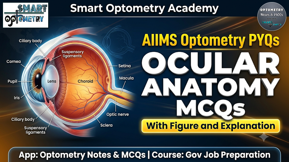
AIIMS Optometry PYQs- Ocular Anatomy
Watch the video explanation, then solve 10 free AIIMS Optometry PYQs on ocular anatomy with detailed answers. 17 bonus MCQs are locked inside the app.
🎥 Watch: AIIMS Optometry Ocular Anatomy PYQs – Full Breakdown
📌 This video covers the same MCQs. After watching, test yourself with the interactive quiz below.
Q1. The cornea is supplied by nerve fibers derived from: [AIIMS Optometry PYQs]
A Trochlear nerve
B Optic nerve
C Trigeminal nerve
D Oculomotor nerve
💡 Explanation (Answer: C)
The cornea is one of the most highly sensitive tissues in the human body, receiving rich sensory innervation. These sensory fibers are derived from the long ciliary nerves, which are branches of the ophthalmic division of the trigeminal nerve (cranial nerve V).
Q2. Which layer of the retina is first encountered by light? [AIIMS Optometry PYQs]
A Inner limiting membrane
B Outer limiting membrane
C Layers of rods and cones
D Ganglion cell layer
💡 Explanation (Answer: A)
Light entering the vitreous chamber must pass through almost all inner retinal layers before reaching the photoreceptors. The inner limiting membrane, formed by the footplates of Muller cells, serves as the anatomical boundary and the first layer encountered by light.
🚀 Govt Job Preparation – Full Course
Gain immediate access to premium learning materials tailored perfectly for upcoming state & central-level Optometry Government Exams. Let’s make learning highly systematic.
Q3. Name the point which has the maximum concentration of cone cells in the retina. [AIIMS Optometry PYQs]
A Fovea centralis
B Fovea
C Optic chiasma
D Blind spot
💡 Explanation (Answer: A)
The fovea centralis is the rod-free central zone of the macula that contains the highest spatial density of cone photoreceptors. This dense packing of cones is essential for achieving peak visual acuity and optimal color discrimination.
Q4. Which of the following is the correct pair regarding extraocular muscle and their primary action? [AIIMS Optometry PYQs]
I. Superior rectus – Elevation
II. Superior oblique – Intorsion
A Only II
B Only I
C Neither I nor II
D Both I and II
💡 Explanation (Answer: D)
The superior rectus muscle primarily elevates the globe when the eye is in an abducted position. The superior oblique muscle operates through the trochlear pulley to primarily produce intorsion of the eye.
📚 Everything You Need for Optometry Exams
Secure high scores in clinical, optical, and anatomical domains. Our mock trials and detailed syllabus-mapped notes keep your preparation organized.
Q5. Which is the longest extraocular muscle in the human eye? [AIIMS Optometry PYQs]
A Superior Rectus
B Inferior Rectus
C Blue dot cataract
D Superior Oblique
💡 Explanation (Answer: D)
The superior oblique muscle is the longest extraocular muscle, extending from its origin at the orbital apex to its insertion on the posterolateral sclera. Its functional path is uniquely lengthened because its tendon must thread through a fibrous pulley called the trochlea before reaching the globe.
Q6. Which layer of the cornea is primarily responsible for its tensile strength? [AIIMS Optometry Previous Year MCQs]
A Epithelium
B Bowman’s membrane
C Stroma
D Descemet’s membrane
💡 Explanation (Answer: C)
The corneal stroma accounts for approximately 90% of the total corneal thickness and is composed of highly organized collagen lamellae. This unique spatial arrangement of parallel collagen fibers provides the cornea with outstanding structural durability and tensile strength.
🚀 Govt Job Preparation – Full Course
Gain immediate access to premium learning materials tailored perfectly for upcoming state & central-level Optometry Government Exams. Let’s make learning highly systematic.
Q7. The axons of which retinal cells converge to form the Optic Nerve? [AIIMS Optometry Previous Year MCQs]
A Photoreceptor cells
B Bipolar cells
C Ganglion cells
D Amacrine cells
💡 Explanation (Answer: C)
Retinal ganglion cells receive visual signals from photoreceptors via bipolar and amacrine cells. Their long, unmyelinated axons course along the nerve fiber layer to converge at the optic disc, where they form the optic nerve.
Q8. The region of the retina where no image is formed and contains no rods or cones is known as: [AIIMS Optometry Previous Year MCQs]
A Choroid
B Blind spot
C Yellow spot
D Cornea
💡 Explanation (Answer: B)
The optic disc, commonly known as the blind spot, is the anatomical site where retinal ganglion cell axons exit the eye to form the optic nerve. Because this region completely lacks photoreceptor cells, it is entirely insensitive to light and cannot form visual images.
📚 Everything You Need for Optometry Exams
Secure high scores in clinical, optical, and anatomical domains. Our mock trials and detailed syllabus-mapped notes keep your preparation organized.
Q9. The choroid layer is full of: [AIIMS Optometry Previous Year MCQs]
A Tubes
B Veins
C Blood vessels
D Muscles
💡 Explanation (Answer: C)
The choroid is a highly vascular, pigmented tissue layer situated between the retina and the sclera. Its extensive network of blood vessels supplies vital oxygen and nutrients to the outer layers of the retina, particularly the photoreceptors.
Q10. The function of the iris is: [AIIMS Optometry Previous Year MCQs]
A Carries visual messages to the brain
B Regulates the amount of light entering the eye
C Converts light into electrical impulses
D Gives central vision
💡 Explanation (Answer: B)
The iris contains two antagonistic muscles, the sphincter pupillae and dilator pupillae, which dynamically adjust the diameter of the pupil. By contracting or relaxing these muscles, the iris regulates the exact volume of ambient light entering the posterior segment of the eye.
🔒 Bonus MCQs – Available in App
Unlock these 17 exclusive premium questions + all 15 clinical subjects in the Govt Job Preparation course.
Q11. The clear layer on the top of the eye is:
A The lens
B The conjunctiva
Q12. The unique structure for each individual is the:
A Conjunctiva
B Iris
Q13. Match the correct pairs: 1. Optic nerve (Carries electrical impulses), 2. Fovea (Sharpest vision), 3. Lens (Helps focus)
A 1a, 2b, 3c
B 1b, 2c, 3a
Q14. The lateral rectus muscle is supplied by which cranial nerve?
A Cranial Nerve IV
B Cranial Nerve VI
Q15. In which portion of the retina is maximal visual acuity present?
A 20 degrees Temporal to fovea
B Fovea
Q16. Which structure in the eye is highly vascular?
A Retina
B Choroid
Q17. How many rectus and oblique muscles are attached to each eye?
A Two, four
B Four, two
Q18. The ring-shaped ciliary body is formed by the extension of the ______ towards the inside of the eye.
A Retina
B Choroid
Q19. Which layer of the eye contains rods and cones?
A Choroid
B Retina
Q20. Which of the following is NOT a type of extraocular muscle?
A Superior rectus
B Medial oblique
Q21. Keratic Precipitates (KPs) are inflammatory deposits typically found on:
A Corneal Epithelium
B Corneal Endothelium
Q22. What is the average axial length of a normal adult human eye?
A 22 mm
B 24 mm
Q23. Which of the following is NOT one of the layers of the ciliary body?
A Sphincter pupillae
B Pigmented epithelium
Q24. Which layer of cornea consists of acellular mass of condensed collagen fibrils?
A Stroma
B Bowman’s membrane
Q25. Which of the following statements is correct regarding the anatomy of sclera?
A Only I
B Both I and II
Q26. The corneal epithelium is resistant to all organisms EXCEPT:
A E. coli
B Listeria
Q27. Which of the following constitutes approximately 70 percent of the dry weight of the human cornea?
A Glycoproteins
B Collagen
🎯 Accelerate Your Optometry Exam Prep
Access high-yield study resources, smart mock tests, and a high-performance community.
🎯 Enroll in Full Govt Job Preparation Course